- HOME
- Department
- Histopathology
Histopathology
(1) Overview
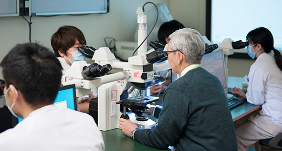
Histopathology is a department that conducts “pathological diagnosis” to observe tissues and cells collected from patients by examination under a microscope and find out what the disease is. There are various examinations to diagnose the disease such as blood test, and urine test. Some diseases such as cancer, we can make a definite diagnosis by examining the tissue under a microscope. Collecting tissues will be done to examine them, using the way called biopsy, before the treatment policy is decided, or during the surgery. The result of pathological diagnosis will promptly be informed to the attending physicians and will be used for deciding the treatment plan.
We have regular conferences with the doctors of each department to share the information and have a discussion about the treatment plan.
(2) Policy
We think that the accuracy and promptness of the diagnosis is the most important task to contribute to the improvement of the quality of the medical service to provide for our patients. We, therefore, manage the quality control of the diagnosis by thoroughly performing double-checks by more than two Pathologists certified by the Japanese Society of Pathology. For the cases which are difficult to diagnose, all of the Pathologists will be involved in the conferences and will seek for the consultation to external specialists if required to keep the accuracy.
When handling with specimens (tissues and cells) collected from patients, we pay close attention to privacy protection so that individuals are not identified.
(3) Our Strengths
When We Make the Cytodiagnosis
For lung cancer and urinary bladder cancer, the cancer cells may be excreted to phlegm or urine. Therefore, when these cancers are suspected, sputum and urine may be collected and a cytodiagnosis may be performed to check for the presence of cancer cells under a microscope.
When lump exists in the breast, we will suck the tissues using a thin needle to make the pathological diagnosis.
For the cervical cancer screening, we will make the scraping cytology by scraping the part of the uterine cervix by a brush.
Purpose of the Biopsy Tissue Diagnosis
When lesions found by various examinations such as ultrasound examination or endoscopy, the biopsy will be performed to confirm the diagnosis and decide the treatment plan. The biopsy is an examination to collect lesions by plunking or cutting off by a scalpel.
We make the pathological diagnosis called as the histological diagnosis using the collected specimens. Based upon the result, we will decide if these are the leisions that will require the surgery, or will be subject to the medical follow up.
The biopsy tissue diagnosis is for the areas reachable by the endoscopy, such as stomach, or large intestine, or lesions reachable by needle, or the incision of the skin.
Rapid Diagnosis During the Surgery
If the biopsy is not feasible before the surgery as the lesions are located in the deep area of the body, we will make the pathological diagnosis by collecting tissues during the surgery. This is called as the rapid diagnosis, which will take ~ 10 minutes and the result will be informed to the surgeon who performs the surgery promptly. Through the rapid diagnosis, the confirmation whether the lesions are completely removed or the diagnosis whether there is metastasis or not can be made so that the final decision of the ongoing surgery plan can be made.